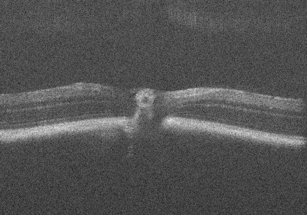
Optical Coherence Tomography: Imaging Mouse Retinal Ganglion Cells In Vivo
$ 25.50 · 4.9 (208) · In stock

Scientific Article | Structural changes in the retina are common manifestations of ophthalmic diseases.

PDF] In vivo imaging and counting of rat retinal ganglion cells using a scanning laser ophthalmoscope.

Optical Coherence Tomography: Imaging Mouse Retinal Ganglion Cells In Vivo. - Abstract - Europe PMC

PDF) In Vivo Imaging of Cx3cr1gfp/gfp Reporter Mice with Spectral-domain Optical Coherence Tomography and Scanning Laser Ophthalmoscopy

All Protocols and Video Articles in JoVE
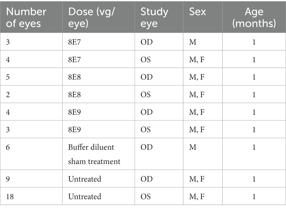
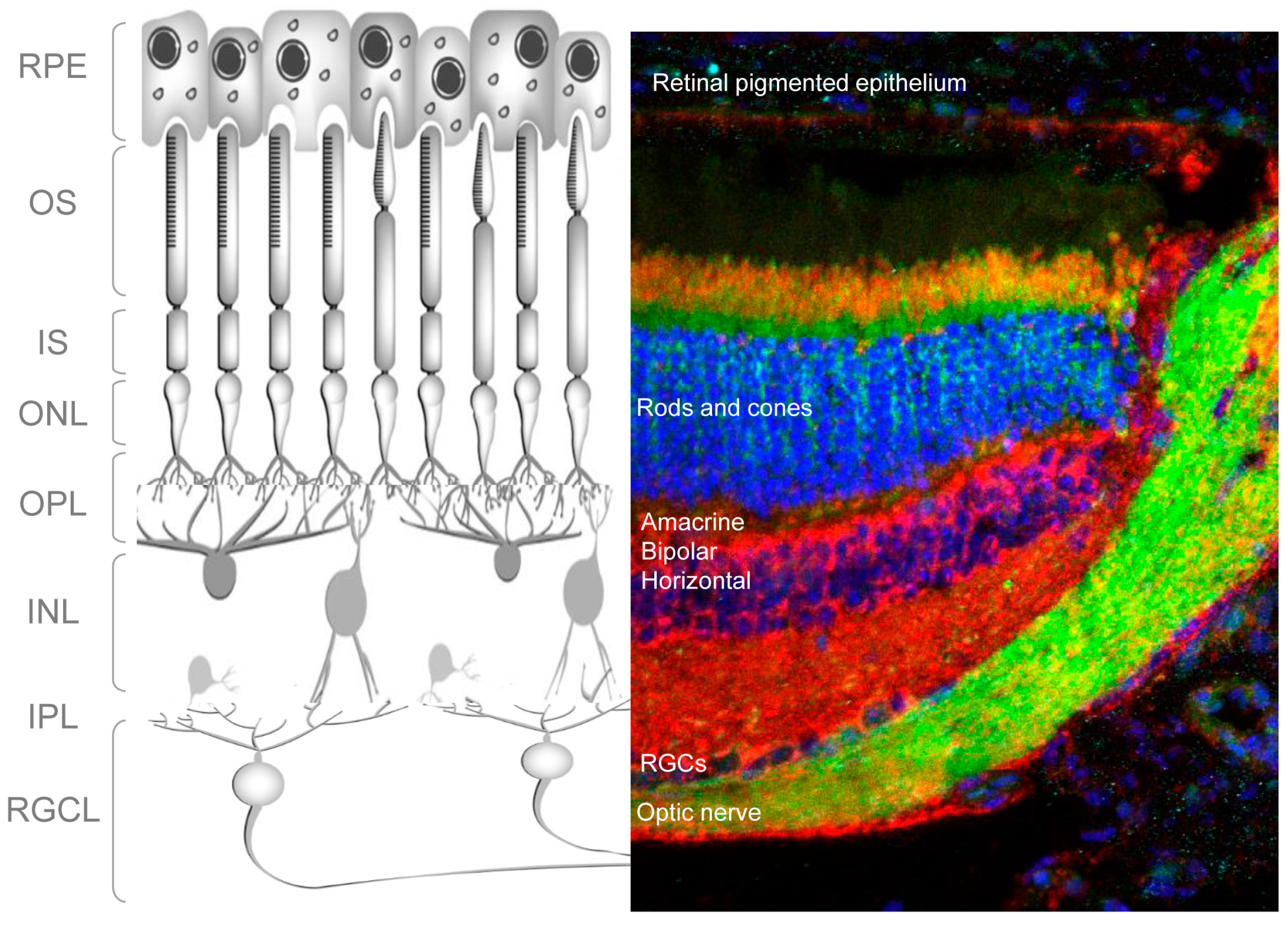
Frontiers The dose-response relationship of subretinal gene therapy with rAAV2tYF-CB-hRS1 in a mouse model of X-linked retinoschisis

Topical photodynamic therapy combined with ablative “light needles” against basal cell carcinoma - ScienceDirect

In vivo imaging of the inner retinal layer structure in mice after eye-opening using visible-light optical coherence tomography - ScienceDirect

Researching Review of Advances in Ophthalmic Optical Imaging Technologies from Several Mouse Retinal Imaging Methods

Choroidal thickness and the retinal ganglion cell complex in
Cells, Free Full-Text

Non-invasive in vivo hyperspectral imaging of the retina for potential biomarker use in Alzheimer's disease

Optical Coherence Tomography: Imaging Mouse Retinal Ganglion Cells In Vivo
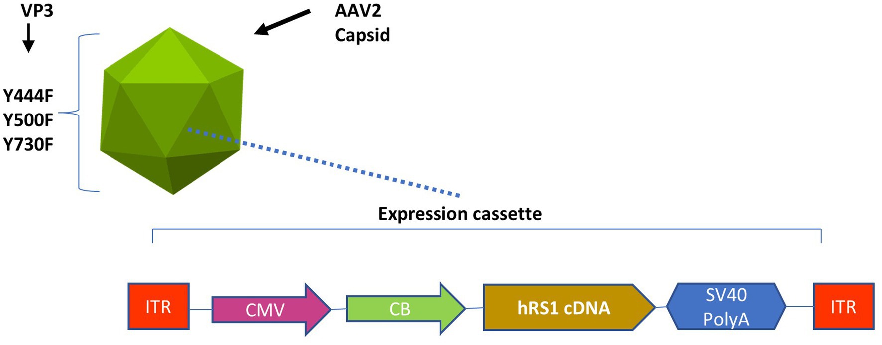
Frontiers The dose-response relationship of subretinal gene therapy with rAAV2tYF-CB-hRS1 in a mouse model of X-linked retinoschisis
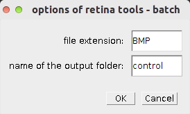
Retina Tool - ImageJ-macros - MRI's Redmine
Retina Tool - ImageJ-macros - MRI's Redmine